Giorgia Baldascino
⏳ 3 min
At the end of September, my time as a research fellow came to an end — a chapter carried out under the guidance of Prof. Veronese and Dr. Moretto.
In truth, this journey had started a year earlier, with my master’s thesis. It has been an intense adventure filled with new challenges, countless lines of code, and brain images to analyze — but also with inspiring encounters with curious and passionate people who have walked this path with me.
The project I worked on is part of iMarkHD, a collaboration with King’s College London focused on finding biomarkers for Huntington’s disease. The goal is to detect signs of the disease even before symptoms appear, so that early interventions could slow — or even stop — its progression.
One of the main tools in this research is PET imaging, which allows us to observe brain activity. However, PET has an important limitation: its low spatial resolution, which causes what’s known as the partial volume effect — a kind of “blurring” that mixes signals from different brain regions. This can lead to underestimating certain measures and reduce the sensitivity of analyses, especially in small regions or those that shrink as the disease progresses, as happens in Huntington’s. Correcting this problem is therefore crucial for obtaining accurate and reliable results.
To tackle this issue, several partial volume correction (PVC) methods have been developed. Some of the most promising ones combine PET images with high-resolution MRI, integrating structural and functional information more effectively.
Despite their potential, only a few PET studies on Huntington’s disease have applied these techniques systematically. Too often, partial volume effects are overlooked or not considered as a source of error. This highlights the need to rigorously evaluate existing methods to ensure more reliable analyses and stronger findings in biomarker research.
That’s exactly where my work comes in. During my thesis, and later throughout my fellowship, I explored the scientific literature, selected publicly available correction algorithms, and applied them to PET data from both Huntington’s patients and healthy volunteers. Three of these methods proved particularly promising, and I focused my analysis on them with the goal of assessing their reliability and contributing to the identification of a reference approach for this type of study.
The results confirmed that correcting for the partial volume effect truly matters. All the tested methods improved the accuracy of PET measurements. Among them, the correction proposed by Zhu et al. turned out to be the most reliable and reproducible, especially in the brain regions most affected by Huntington’s disease. This strengthens the idea that PVC is a key tool for making PET a robust biomarker in the study of this condition.
Working on this project allowed me to develop new skills in software development, data analysis, and brain imaging — while also applying and expanding the knowledge I gained during my studies. Collaborating with such curious and passionate researchers has been an incredibly inspiring experience.
This was not just an academic exercise, but a project with real potential impact on Huntington’s disease research and, more broadly, on the use of PET in studying neurodegenerative disorders. Knowing that I’ve contributed — even in a small way — to a field where there is still so much to discover makes me proud of this journey.
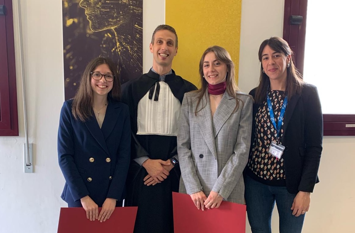
A personal highlight was receiving the “Silvio Cavalcanti” Award from the University of Bologna, granted by the Italian National Group of Bioengineering, for the thesis project that started it all — an unexpected recognition that made this path even more special.
I’m deeply grateful to my supervisors, colleagues, and everyone I’ve met along the way for everything they’ve shared with me and for helping me grow, both professionally and personally.
I hope that the work I’ve done will be useful not only for the iMarkHD project but also for future studies, contributing — even just a little — to advancing research in this field.
• • •
Alla fine di settembre si è concluso il mio percorso come borsista di ricerca, svolto sotto la supervisione del prof. Veronese e della dott.ssa Moretto.
In realtà questa esperienza era iniziata già un anno fa, con la mia tesi magistrale. È stato un viaggio intenso, fatto di nuove sfide, tante righe di codice e immagini cerebrali da analizzare, ma anche di incontri con persone curiose e appassionate che mi hanno accompagnata lungo la strada.
Il progetto a cui ho lavorato si inserisce nello studio iMarkHD, una collaborazione con il King’s College di Londra dedicata alla ricerca di biomarcatori per la malattia di Huntington. L’obiettivo è quello di individuare segnali della malattia ancora prima della comparsa dei sintomi, così da poter intervenire tempestivamente per rallentarne – o addirittura fermarne – la progressione.
Uno degli strumenti principali per raggiungere questo obiettivo è la PET, una tecnica di neuroimaging che ci permette di osservare l’attività cerebrale. La PET, però, ha un limite importante: la sua bassa risoluzione spaziale, che provoca il cosiddetto partial volume effect, un effetto di “sfocatura” che mescola i segnali provenienti da regioni diverse del cervello. Questo porta a sottostimare alcune misure e riduce la sensibilità delle analisi, soprattutto in regioni molto piccole o che si riducono con la progressione della malattia, come avviene nell’Huntington. Correggere questo problema è quindi fondamentale per rendere i risultati più precisi e affidabili.
Per ridurre questo problema, esistono diversi metodi di correzione del volume parziale (PVC). Alcuni, tra i più promettenti, combinano immagini PET con immagini di risonanza magnetica ad alta risoluzione, così da integrare al meglio informazioni strutturali e funzionali.
Nonostante la loro importanza, però, solo pochi studi PET sull’Huntington hanno applicato queste tecniche in modo sistematico. Spesso i partial volume effects vengono ignorati o non considerati come una possibile fonte di errore. Questo evidenzia l’urgenza di valutare in maniera più rigorosa i metodi disponibili, così da garantire analisi più affidabili e risultati più solidi nella ricerca sui biomarcatori.
Ed è proprio qui che si inserisce il mio lavoro. Durante la tesi, e poi con la borsa di ricerca, mi sono occupata di esplorare la letteratura scientifica, selezionare algoritmi di correzione disponibili pubblicamente e applicarli ai dati PET di pazienti con Huntington e di soggetti sani. Tre di questi si sono rivelati particolarmente promettenti ed è su di loro che ho concentrato la mia analisi, con l’obiettivo di valutarne l’affidabilità e contribuire a individuare un metodo di riferimento per questo tipo di studi.
I risultati hanno confermato che correggere il partial volume effect è fondamentale. Tutti i metodi testati hanno migliorato la precisione delle misure PET. Tra questi, la correzione proposta da Zhu et al. si è dimostrata la più affidabile e riproducibile, soprattutto nelle regioni cerebrali più colpite dalla malattia di Huntington. Questo rafforza l’idea che la PVC sia uno strumento cruciale per rendere la PET un biomarcatore davvero solido nello studio della malattia.
Svolgere questo progetto mi ha permesso di acquisire nuove competenze in ambito software, analisi di dati e immagini cerebrali, oltre a consolidare e applicare alcune delle conoscenze maturate durante il mio percorso di studi. Poter lavorare a contatto con persone così curiose e appassionate di ricerca in questo ambito è stato davvero stimolante
Non è stato solo un esercizio accademico, ma un lavoro con un potenziale impatto concreto sulla ricerca sulla malattia di Huntington e, più in generale, sull’uso della PET nello studio delle neurodegenerazioni. Sapere di aver contribuito, anche in piccolo, a un ambito dove c’è ancora tanto da fare mi rende orgogliosa e fiera del mio lavoro.
Un piccolo grande motivo di orgoglio personale è che, grazie al progetto di tesi da cui tutto è iniziato, ho ricevuto il Premio di laurea “Silvio Cavalcanti” dell’Università di Bologna, erogato dal Gruppo Nazionale di Bioingegneria. Una sorpresa che ha reso questo percorso ancora più speciale.
Sono profondamente grata ai miei supervisori, ai colleghi e a tutte le persone che ho incontrato durante questo percorso, per tutto ciò che mi hanno trasmesso e che mi ha permesso di crescere sia professionalmente sia personalmente.
Spero che il lavoro svolto possa essere utile non solo al progetto iMarkHD, ma anche ad altri studi futuri, contribuendo – anche solo un po’ – a migliorare la ricerca in questo ambito.
Leave a Reply